Biopsy

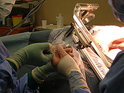
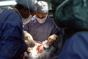
A biopsy is a medical test involving the removal of cells or tissues for examination. It is the medical removal of tissue from a living subject to determine the presence or extent of a disease. The tissue is generally examined under a microscope by a pathologist, and can also be analyzed chemically. When an entire lump or suspicious area is removed, the procedure is called an excisional biopsy. When only a sample of tissue is removed with preservation of the histological architecture of the tissue’s cells, the procedure is called an incisional biopsy or core biopsy. When a sample of tissue or fluid is removed with a needle in such a way that cells are removed without preserving the histological architecture of the tissue cells, the procedure is called a needle aspiration biopsy.
Contents |
Etymology
Biopsy is of Greek origin, coming from the words bio, meaning life, and opsia, meaning to see.
Breast biopsy
Several methods for a breast biopsy now exist. The most appropriate method of biopsy for a patient depends upon a variety of factors, including the size, location, appearance and characteristics of the abnormality.
Fine needle aspiration
Fine needle aspiration (FNA) is a percutaneous ("through the skin") procedure that uses a fine needle and a syringe to sample fluid from a breast cyst or remove clusters of cells from a solid mass. With FNA, the cellular material taken from the breast is usually sent to the pathology laboratory for analysis. A technique similar to FNA can also be used by the radiologist or surgeon to drain fluid from a benign cyst. This procedure is called cyst aspiration. A fine needle aspiration procedure is generally almost painless and takes only a few minutes to perform. Technically, this FNA procedure is not a biopsy as the material retrieved is made of fluid coming either from a cyst or from the intercellular space and a few cells, when a biopsy is bringing back a piece of tissue where the architecture of the tissue is preserved.
Core needle biopsy

A core needle biopsy is a procedure that removes small but solid samples of tissue using a hollow "core" needle. For palpable (“able to be felt”) lesions, the physician fixes the lesion with one hand and performs a freehand needle biopsy with the other. In case of non-palpable lesions stereotactic mammography, or ultrasound, or PEM guidance is used. With stereotactic mammography it is possible to pinpoint the exact location of a mass based on images taken from two different angles of the x-ray machine. With ultrasound, the radiologist or surgeon can watch the needle on the ultrasound monitor to help guide it to the area of concern. With PEM (positron emission mammagraphy), the lesion is targeted in 3D based on a positron emission tomography (PET) image of the breast. The needle used during core needle biopsy is larger than the needle used with FNA. The core biopsy needle also has a special cutting edge allowing removal of a bigger sample of tissue. With core needle biopsy a relatively large sample can be removed through a small single incision in the skin. Typically, the breast area is first locally anesthetized with a small amount of anesthetic fluid. Then, the needle is placed into the breast. As with FNA, the radiologist or surgeon will guide the needle into the area of concern by palpating the lump. If the lesion can’t be felt the core needle biopsy is performed under image-guidance using either stereotactic mammography, ultrasound or even magnetic resonance imaging (MRI). A core needle biopsy procedure takes a few minutes to perform and is almost painless.
Vacuum assisted biopsy
Vacuum assisted biopsy is a version of core needle biopsy using a vacuum technique to assist the collection of the tissue sample. The needle normally has a lateral (“from the side”) opening and can be rotated allowing multiple samples to be collected through a single skin incision. The Vacuum assisted biopsy procedure is similar to normal core needle biopsy. The vacuum assisted biopsy category also includes automated rotational core devices.[1]
Direct & Frontal Biopsy
Recent innovations in tissue acquisition for the human breast have led to the development of unique direct frontal systems. Efficacy is considered optimal if the diagnosis by transcutaneous biopsy is identical to the surgical specimen in case of malignancy or in line with clinical follow-up when benign. The direct and frontal biopsy systems are safe for the patient, and the procedure can even be considered relatively painless. The quality of the sample is sufficient for research on molecular biology. The direct frontal tissue-acquisition systems can be added to the list of safe and efficient macrobiopsy methods to be used in the early detection of breast cancer.
Open surgical biopsy

Open surgical biopsy means that a large mass or lump is removed during a surgical procedure. Surgical biopsy requires an approximately 3 to 5 centimeters incision and is normally performed in an operating room in sterile conditions. Open surgical biopsy in some cases can be performed with local anesthesia but in most cases general anesthesia may be necessary. Ten years ago, most breast biopsies were open surgical procedures. Today most patients are candidates for less invasive biopsy procedures such as core needle biopsy. Depending on the location of the lesion to be biopsied, a radiologist will often perform needle localization beforehand to guide the surgeon to the site being biopsied.
Skin biopsy
Multiple methods for skin biopsy exist. Each has its own limitation and problems. Most are done under local anesthesia in a doctor's office. The result is very dependent on the clinical history presented to the pathologist, and also the method utilized. A shave biopsy is absolutely useless in diagnosising vasculitis, whereas an excisional biopsy might be excessive in diagnosing a possible basal cell carcinoma.
Shave biopsy
This is done with either a small scalpel blade, a curved razor blade, or a broken piece of "safety" razor. The technique is very much user skill dependent, as some surgeons can remove a small fragment of skin with minimal blemish using any one of the above tools, while other have great difficulty securing the devices. Ideally, the razor will shave only a small fragment of protruding tumor and leaving the skin relatively flat after the procedure. Hemostasis is obtained using light electrocautery, Monsel solution, or aluminum chloride. This is the ideal method of diagnosis for basal cell cancer. It can be used to diagnose squamous cell carcinoma and melanoma-in-situ, however, the doctor's understanding of the growth of these last two cancers should be considered before one uses the shave method. The punch or incisional method is better for the latter two cancers as false negative is less likely to occur (i.e. calling a squamous cell cancer an actinic keratosis or keratinous debris). Hemostasis for the shave technique can be difficult if one relied on electrocautery alone. A small "shave" biopsy often ends up being a large burn defect when the surgeon tries to control the bleeding with electrocautery alone. Pressure dressing or chemical astringent can help in hemostasis in patients taking anticoagulants.
Punch biopsy
This is done with a round shaped knife ranging in size from 1mm to 8 mm. Some punch biopsies are shaped like an ellipse, although one can accomplish the same desired shape with a standard scalpel. The 1 mm and 1.5 mm punch are ideal for locations where cosmetic appearance is difficult to accomplish with the shave method. Minimal bleeding is noted with the 1 mm punch, and often the wound is left to heal without stitching for the smaller punch biopsies. Disadvantage of the 1 mm punch is that the tissue obtained is almost impossible to see at times due to small size, and the 1.5 mm biopsy is preferred in most cases. The common punch size use to diagnose most inflammatory skin condition is the 3.5 or 4 mm punch. Ideally, the punch biopsy include the full thickness skin and subcutanous fat in the diagnosis of skin diseases. The punch biopsy is preferred over the shave biopsy for the diagnosis of squamous cell carcinoma and for melanomas. One or two sutures are required to close most punch biopsies with the exception of the smallest punches. Two "dog ear" defects can result in punch biopsies much larger than 5 mm, thus an incisional biopsy is preferred on larger lesions.
Incisional biopsy
When a cut is made through the entire dermis down to the subcutanous fat. A punch biopsy is essentially an incisional biopsy, except it is round rather than elliptical as in most incisional biopsies done with a scalpel. Incisional biopsies can include the whole lesion (excisional), part of a lesion, or part of the affected skin plus part of the normal skin (to show the interface between normal and abnormal skin). Incisional biopsy often yield better diagnosis for deep pannicular skin diseases and more subcutanous tissue can be obtained than a punch biopsy. Long and thin deep incisional biopsy are excellent on the lower extremities as they allow a large amount of tissue to be harvested with minimal tension on the surgical wound. Advantage of the incisional biopsy over the punch method is that hemostasis can be done more easily due to better visualization. Dog ear defects are rarely seen in incisional biopsies with length at least twice as long as the width.
Excisional biopsy
This is essentially the same as incision biopsy, except the entire lesion or tumor is included. This is the ideal method of diagnosis of small melanomas (when performed as an excision). Ideally, an entire melanoma should be submitted for diagnosis if it can be done safely and cosmetically. This "excisional" biopsy is often done with a narrow margin to make sure the deepest thickness of the melanoma is given before prognosis is decided. However, as many melanoma-in-situs are large and on the face, a physician often chose to do multiple small punch biopsies before committing to a large excision for diagnostic purpose alone. Many prefer the small punch method for initial diagnostic value before resorting to the excisional biopsy. An initial small punch biopsy of a melanoma might say "severe cellular atypia, recommend wider excision". At this point, the clinician can be confident that an excisional biopsy can be performed without risking committing a "false positive" clinical diagnosis.
Curettage biopsy
This can be done on the surface of tumors or on small epidermal lesions with minimal to no topical anesthetic using a round curette blade. Diagnosis of basal cell cancer can be made with some limitation, as morphology of the tumor is often disrupted. The pathologist must be informed about the type of anesthetic used, as topical anesthetic can cause artifact in the epidermal cells.
Fine needle aspirate
This is done with the rapid stabbing motion of the hand guiding a needle tipped syringe and the rapid sucking motion applied to the syringe. It is a method used to diagnose tumor deep in the skin or lymphnodes under the skin. The cellular aspirate is mounted on a glass slide and immediate diagnosis can be made with proper staining or submitted to a laboratory for final diagnosis. A fine needle aspirate can be done with simply a large bore needle and a small syringe (1 cc) that can generate rapid changes in suction pressure. Fine needle aspirate can be used to distinguish a cystic lesion from a lipoma. Both the surgeon and the pathologist must be familiar with the method of procuring, fixing, and reading of the slide. Many center have dedicated team used in the harvest of fine needle aspirate.
"Scoop", "scallop", or "shave" excision
A trend has occurred in dermatology over the last 10 years with the advocacy of a deep shave excision of a pigmented lesion. An author published the result of this method and advocated it as better than standard excision and less time consuming. The added economic benefit is that many surgeons bill the procedure as an excision, rather than a shave biopsy. This save the added time for hemostasis, instruments, and suture cost. The great disadvantage, seen years later is the numerous scallop scars, and a very difficult to deal with lesion called a "recurrent melanocytic nevus". What has happened is that many "shave" excisions does not adequately penetrate the dermis or subcutanous fat enough to include the entire melanocytic lesion. Residual melanocytes regrow into the scar. The combination of scarring, inflammation, blood vessels, and atypical pigmented streaks seen in these recurrent nevus gives the perfect dermatoscopic picture of a melanoma. When a second physicians re-examine the patient, he or she has no choice but to recommend the reexcision of the scar. If one does not have access to the original pathology report, it is impossible to tell a recurring nevus from a severely dysplastic nevus or a melanoma. As the procedure is widely practiced, it is not unusual to see a patient with dozens of scallop scars, with as many as 20% of the scar showing residual pigmentation. The second issue with the shave excision is fat herniation, iatrogenic anetoderma, and hypertrophic scarring. As the deep shave excision either completely remove the full thickness of the dermis or greatly diminishing the dermal thickness, subcutanous fat can herniate outward or pucker the skin out in an unattractive way. In areas prone to friction, this can result in pain, itching, or hypertrophic scarring.
History
One of the earliest diagnostic biopsies was developed by the Arab physician Abulcasim (1013-1107 AD). A needle was used to puncture a goiter, and the material issuing was characterized.[6]
Cancer
When cancer is suspected, a variety of biopsy techniques can be applied. An excisional biopsy is an attempt to remove an entire lesion. When the specimen is evaluated, in addition to diagnosis, the amount of uninvolved tissue around the lesion, the surgical margin of the specimen is examined to see if the disease has spread beyond the area biopsied. "Clear margins" or "negative margins" means that no disease was found at the edges of the biopsy specimen. "Positive margins" means that disease was found, and a wider excision may be needed, depending on the diagnosis. When intact removal is not indicated for a variety of reasons, a wedge of tissue may be taken in an incisional biopsy. In some cases, a sample can be collected by devices that "bite" a sample. A variety of sizes of needle can collect tissue in the lumen (‘’core biopsy’’). Smaller diameter needles collect cells and cell clusters, fine needle aspiration biopsy.[7] Pathologic examination of a biopsy can determine whether a lesion is benign or malignant, and can help differentiate between different types of cancer. In contrast to a biopsy that merely samples a lesion, a larger excisional specimen called a resection may come to a pathologist, typically from a surgeon attempting to eradicate a known lesion from a patient. For example, a pathologist would examine a mastectomy specimen, even if a previous nonexcisional breast biopsy had already established the diagnosis of breast cancer. Examination of the full mastectomy specimen would confirm the exact nature of the cancer (subclassification of tumor and histologic "grading") and reveal the extent of its spread (pathologic "staging").
Precancerous conditions
For easily detected and accessed sites, any suspicious lesions may be assessed. Originally, this was skin or superficial masses. X-ray, then later CT, MRI, and ultrasound along with endoscopy extended the range.
Inflammatory conditions
A biopsy of the temporal arteries is often performed for suspected vasculitis. In inflammatory bowel disease (Crohn's disease and ulcerative colitis), frequent biopsies are taken to assess the activity of disease and to assess changes that precede malignancy.[8]
Biopsy specimens are often taken from part of a lesion when the cause of a disease is uncertain or its extent or exact character is in doubt. Vasculitis, for instance, is usually diagnosed on biopsy.
Kidney disease
Biopsy and fluorescence microscopy are key in the diagnosis of alterations of renal function. The immunofluorescence plays vital role in the diagnosis of Crescentic glomerulonephritis.
Infectious disease
Lymph node enlargement may be due to a variety of infectious or autoimmune diseases.
Metabolic disease
Some conditions affect the whole body, but certain sites are selectively biopsied because they are easily accessed. Amyloidosis is a condition where degraded proteins accumulate in body tissues. In order to make the diagnosis, the gingival.
Transplantation
Biopsies of transplanted organs are performed in order to determine that they are not being rejected or that the disease that necessitated transplant has not recurred.
Fertility
A testicular biopsy is used for evaluating the fertility of men and find out the cause of a possible infertility, e.g. when sperm quality is low, but hormone levels still are within normal ranges.[9]
Commonly biopsied sites
Bone marrow
Since blood cells are formed in the bone marrow, a bone marrow biopsy is employed in the diagnosis of abnormalities of blood cells when the diagnosis cannot be made from the peripheral blood alone. In malignancies of blood cells (leukemia and lymphoma) a bone marrow biopsy is used in staging the disease. The procedure involves taking a core of trabecular bone using a trephine, and then aspirating material.
Gastrointestinal tract
Flexible endoscopy enables access to the upper and lower gastrointestinal tract, such that biopsy of the esophagus, stomach and duodenum via the mouth and the rectum, colon and terminal ileum are commonplace. A variety of biopsy instruments may be introduced through the endoscope and the visualized site biopsied. Until recently, the majority of the small intestine could not be visualized for biopsy. The double-ballon “push-pull” technique allows visualization and biopsy of the entire gastrointestinal tract.[10].
Needle core biopsies or aspirates of the pancreas may be made through the duodenum or stomach.[11]
Lung
Biopsies of the lung can be performed in a variety of ways depending on the location.
Liver
In hepatitis, most biopsies are not used for diagnosis, which can be made by other means. Rather, it is used to determine response to therapy which can be assessed by reduction of inflammation and progression of disease by the degree of fibrosis or, ultimately, cirrhosis.
In Wilson's disease, the biopsy is used to determine the quantitative copper level.
Analysis of biopsied material
After the biopsy is performed, the sample of tissue that was removed from the patient is sent to the pathology laboratory. A pathologist is a physician who specializes in diagnosing diseases (such as cancer) by examining tissue under a microscope. When the laboratory (see Histology) receives the biopsy sample, the tissue is processed and an extremely thin slice of tissue is removed from the sample and attached to a glass slide. Any remaining tissue is saved for use in later studies, if required. The slide with the tissue attached is treated with dyes that stain the tissue, which allows the individual cells in the tissue to be seen more clearly. The slide is then given to the pathologist, who examines the tissue under a microscope, looking for any abnormal findings. The pathologist then prepares a report that lists any abnormal or important findings from the biopsy. This report is sent to the physician who originally performed the biopsy on the patient.
See also
- Bone marrow examination
- Endometrial biopsy
- Lymph node biopsy
- Skin biopsy
References
- ↑ Coding Breast Diseases and Surgery
- ↑ Efficacy and safety of direct and frontal macrobiopsies in breast cancer Ann Cornelisa, Marcel Verjansa, Thierry Van den Boscha, Katrien Wouters, Johan Van Robaeysc, Jaak Ph. Janssensd and Working Group on Hormone Dependent Cancers, The European Cancer Prevention Organization
- ↑ High-Precision Direct and Frontal Breast Biopsy to Assure Adequate Surgical Margin Interpretation; Jaak Janssens, MD, PhD; Ruediger Schulz-Wendtland, MD, PhD; Luc Rotenberg, MD; John-Paul Bogers, MD, PhD 1. University Hasselt, Hasselt, Belgium; 2. University of Erlangen, Erlangen, Germany; 3. Hôpital Hartman, Paris, France; 4. University of Antwerp, Antwerp, Belgium
- ↑ www.medinvents.com
- ↑ European Journal of Cancer Prevention
- ↑ Anderson, J. B., Webb, A.J.: Fine-Needle Aspiration Biopsy and the Diagnosis of Thyroid Cancer. British Journal of Surgery 74:292-6, 1987
- ↑ Sausville, Edward A. and Longo, Dan L.: Principles of Cancer Treatment: Surgery, Chemotherapy, and Biologic Therapy in Harrison's Principles of Internal Medicine, 16th Ed. Kaspar, Dennis L. et al., editors. p.446 (2005)
- ↑ Friedman, S. and Blumberg, R.S.: Inflammatory Bowel Disease in Harrison's Principles of Internal Medicine, 16th Ed. Kaspar, Dennis L. et al., editors. pp. 1176-1789 (2005)
- ↑ Mens health - Testicular Biopsy
- ↑ Saibeni, S., Rondonotti, E., Iozzelli, A., Spina, L., Tontini, G.E., Cavallaro, F., Ciscato, C., de Franchis, R., Sardanelli, F., Vecchi, M.: Imaging of the Small Bowel in Crohn's Disease: A Review of Old and New Techniques World Journal of Gastroenterology 13(24): 3279-87, 2007
- ↑ Iglesias-Garcia, J., Dominguez-Munoz, E., Lozano-Leon, A., Abdulkader, I., Larino-Noia, J., Antunez, J., Forteza, J.: Impact of Endoscopic Ultrasound-Guided Fine Needle Biopsy for Diagnosis of Pancreatic Masses. World Journal of Gastroenterology 13(2): 289-93, 2007
External links
- Mybiopsyinfo.com - What is a biopsy? How is a biopsy examination performed? This website gives you answers to these and many other questions.
- MyBiopsy.org - Information about biopsy results for patients. This site is created by pathologists, the physicians who diagnose cancer and other diseases by looking at biopsies under a microscope.
- RadiologyInfo - The radiology information resource for patients: Biopsy
- Fine needle aspiration biopsy on Wikisurgery
- Core needle (Trucut) biopsy on Wikisurgery